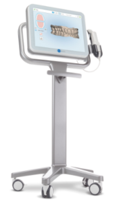
The 3D iTero® Scanner
Your very own digital dental smile design
At Malmin we believe in combining our expertise with newest technology and we’re proud that most of our clinics now offer the new digital state-of-the-art iTero® scanning machines.
The technology enables dental clinicians to provide a realistic digital image of your teeth using ClinCheck 3D® technology. The 3D iTero® Scanner is one of a new breed of technology that is revolutionising dentistry allowing you to have your very own digital dental smile design.
The magic of iTero® scanning lies in providing dental care professionals with more streamlined and efficient practice, supporting diagnostic and treatment planning needs for optimal patient care. The iTero® technology also allows us to create an accurate visual simulation of what your teeth would look like following treatment.
How does it work?
iTero® works by capturing thousands of images of the inside of the mouth and then assembling them into a clinically accurate 3D digital dental smile design of the mouth, teeth, and gums.
Those are then further analysed and inspected in greater detail allowing our clinicians to create the best and most accurate treatment plan for you.

Frequently Asked Questions
How long does it take to do an iTero scan?
Does the iTero scanner use radiation?
The iTero® Scanner is completely radiation-free, which makes it safe for pregnant women.
Is iTero a laser?
The scanning unit emits red laser light (680 nm – Class 1) and white LED light in order to record topographical images of the teeth and oral tissue.

